|
|
|
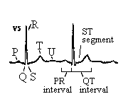
Large EKG grids represent 0.20 seconds and small EKG grids
represent 0.04 seconds,
therefore 5 large squares represent 1 second.
Triplicates: 300, 150, 100, 75, 60, 50, 43, 37, 33, 30
 |
Normal Sinus Rhythm
Rate: 60 - 100; PR interval: 0.12
- 0.20 seconds; QRS interval: 0.04 - 0.12 seconds
|
| Sinus Tachycardia: | Rate: 101 - 160 |
| Sinus Bradycardia: | Rate: 40 - 59 |
| Sinus Arrhythmia: | Irregular; difference between shortest R-R interval and longest R-R interval is at least .12 sec. |
| Sinus Exit Block: | Irregular; length of the pause is the same as 1 cycle, or multiples of cycles; the same rate resumes after the pause; measure the P - P interval |
| Sinus Arrest: | Irregular; the length of the pause is not a multiple of the normal cycle; the rate differs after the pause; measure the P - P interval; document the length of the pause |
| Wandering Atrial Pacemaker: | Irregular; P waves vary; PR interval varies, rate ? 100 |
| Multifocal (Chaotic) Atrial Tachycardia: | Rate: 101 - 250; irregular; P waves of several different shapes, ifpresent, with varying PR intervals |
| Atrial Tachycardia: | Rate:150 - 250; regular; P waves are all the same (if no P waves present, then referred to as Supraventricular Tachycardia) |
| Paroxysmal Atrial Tachycardia: | Rate: 150 - 250; abrupt onset and termination (if no P waves present, then Paroxysmal Supraventricular Tachycardia) |
| Premature Atrial Complexes: | Premature; incomplete compensatory pause; P waves present, but different, or hidden in the T waves |
| Atrial Flutter: | Saw tooth or picket fence pattern P waves; regular;
variable block may
be irregular; document the conduction rate |
| Controlled Atrial Ventricular Fibrillation: | rate: 70 - 110; irregular; no discernible P waves |
| Uncontrolled Atrial Ventricular Fibrillation: | rate: 110 - 220; irregular; no discernible P waves |
| Junctional Rhythm: | Rate: 40 - 60; regular; inverted or biphasic P waves before, during (not visible), or after the QRS |
| Accelerated Junctional Rate: | 61 -100; regular; inverted or biphasic P waves before, during Rhythm: (not visible), or after the QRS |
|
Junctional Tachycardia: |
Rate: 101 - 180; regular; inverted or biphasic P waves before, during (not visible), or after the QRS |
| Premature Junctional Complexes: |
Premature; incomplete compensatory pause; inverted or biphasic P waves before, during (not visible), or after the QRS |
| Ventricular Rhythm: | Rate: 20 - 40; P waves may be present; wide QRS; usually regular, but may be irregular |
| Idioventricular Rhythm: | Rate: 20 - 40; absent P waves; wide QRS; usually regular, but may be irregular |
| Accelerated Idioventricular Rhythm: | Rate: 41 - 99; absent P waves; wide QRS; usually regular, but may be irregular |
| Ventricular Tachycardia: | Rate: 101 - 200+; QRS wide and bizarre; same configuration as PVC; usually regular but may be irregular; 3 or more PVCs in a row |
| Coarse Ventricular Fibrillation: | Rapid, irregular, wide QRS without specific pattern; larger undulations |
| Fine Ventricular Fibrillation: | Rapid irregular QRS without specific pattern; smaller undulations |
| Ventricular Asystole: | Straight or wavy line; no QRS; may be P waves |
| Torsades de Pointes: | Rate: 150 - 300; may be initiated by a PVC
occuring on a prolonged QT interval;
R - R interval is irregular; has spindle effect |
|
Premature Ventricular Complexes: |
Premature; wide and bizarre QRS; T wave deflection is opposite that : of the complex; pause following the PVC is fully compensatory; (R - PVC - R = R - R - R) |
| First Degree Block: | 1 P wave for each QRS complex; PR interval is greater than 0.20 seconds; normal QRS; document the underlying rhythm |
|
Second Degree AV Block |
More P Waves than QRS complexes; PR interval increases Type I progressively until a P is blocked; group beating (Mobitz I or Wenckebach) |
|
Second Degree AV Block - |
More P waves than QRS complexes; PR interval is normal or prolonged, Type II but constant; note the conduction ratio of P:QRS and document (Mobitz II): |
| Third Degree Block | More P waves than QRS complexes; P - P interval is regular; R - R (Complete interval is usually regular, but may be irregular; P waves and QRS Heart Block): complexes are not related to one another; complete AV dissociation |
When analyzing the 12 lead, use a systematic approach:
1) Rate (any lead),
2) Rhythm (lead II),
3) Intervals (PR, QRS, QT),
4) Axis (leads I & AVF),
5) Hypertrophy (lead V1 & V5),
6) Infarct (QRST changes).
| Normal Axis: | positive QRS in both leads I and AVF |
| Left Axis Deviation: | positive QRS in lead I, but negative QRS in lead AVF |
| Right Axis Deviation: | negative QRS in lead I, but positive QRS in lead AVF |
|
Extreme Right Axis Deviation: |
negative QRS in both leads I and AVF |
| Right Atrial Enlargement: | larger initial component of a biphasic P wave in lead V1 or P wave > 2.5 mm in height in any limb lead |
| Left Atrial Enlargement: | wide or ģMī shaped P wave in lead II or larger terminal component of a biphasic P wave in lead V1 |
|
Right Ventricular Hypertrophy: |
R:S ratio > 1 in V1 (R wave > S wave in V1) |
|
Left Ventricular Hypertrophy: |
the depth of the S wave in lead V1 + the height of the R wave in lead V5 > 35 mm (if patient is * age 35) |
| Complete RBBB: | wide QRS, notched R (RRķ) in leads V1 or V2 |
| Incomplete RBBB: | normal QRS, notched R (RRķ) in leads V1 or V2 |
| Complete LBBB: | wide QRS, notched R (RRķ) in leads V5 or V6 |
| Incomplete LBBB: | normal QRS, notched R (RRķ) in leads V5 or V6 |
| Left Anterior Hemiblock: | normal QRS, left axis deviation, Q1 - S3 (Q wave in lead I, S wave in lead III) |
| Left Posterior Hemiblock: | normal QRS, right axis deviation, S1 - Q3 (S wave in lead I, Q wave in lead III) |
|
RBBB + Left Anterior Hemiblock: |
wide QRS, notched R (RRķ) in lead V1, left axis deviation |
|
RBBB + Left Posterior Hemiblock: |
wide QRS, notched R (RRķ) in lead V1, right axis deviation |
The Classic Triad of EKG Characteristics of Myocardial Infarction
ST Segment Elevation - concave down (the earliest change)
at least 2 mm of elevation in 2 contiguous pre-cordial
(chest) leads
at least 1 mm of elevation in 2 contiguous frontal
(limb) leads
T wave Inversion especially in leads V2 - V6
Presence of Q waves
at least .04 sec. wide, depth at least 25% of R wave
or
one-third of the entire QRS amplitude
| Inferior MI | QRST changes in leads II, III, and AVF |
| Anterior MI | QRST changes in leads V1, V2, and V3 |
| Lateral MI | QRST changes in leads I & AVL or V5 & V6 |
| Posterior MI | QRST changes in leads V1 & V2 (in this case look for large R waves and often associated ST segment depression)with Inferior MI |